

Intraoral scanning
A digital model of your mouth.
An intraoral scanner records teeth and oral structures without relying only on conventional impressions. The resulting model supports communication, planning and restorative design.
Integrated digital dentistry
We combine 3D imaging, digital records and CAD/CAM manufacturing to study each case, explain its stages and coordinate personalized treatment.
From diagnosis to restoration
Each tool contributes different information. The appropriate workflow depends on your needs, the available records and findings from the clinical evaluation.
CBCT imaging to study anatomical structures when indicated.
Digital recording of teeth and oral structures.
Recording the spatial relationship between implants.
Integrating records to study position and sequence.
Transferring the digital plan to the procedure when appropriate.
Digital design and milling of the planned restoration.
01 · CBCT imaging
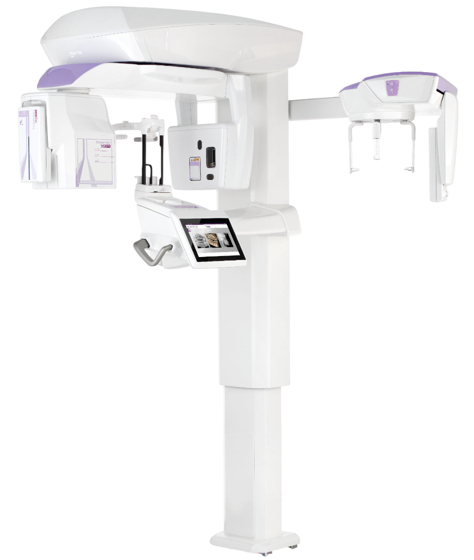
CBCT imaging makes it possible to view teeth, bone and important anatomical structures in three dimensions. This information may support implant evaluation and planning when the study is indicated.
The necessary study and its interpretation must be determined during the clinical evaluation.
02 · Digital capture
Intraoral scanning and photogrammetry help bring clinical information into the digital workflow to coordinate planning and the restorative phase.
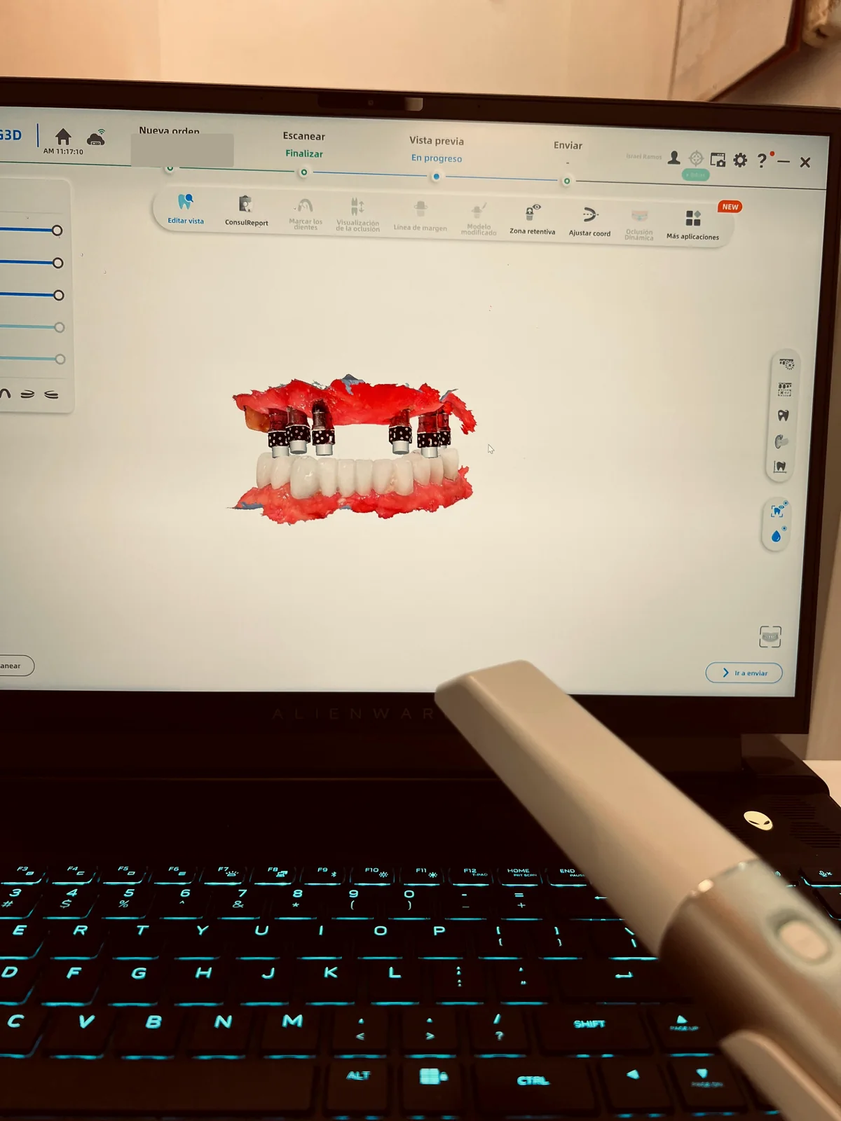
Intraoral scanning
An intraoral scanner records teeth and oral structures without relying only on conventional impressions. The resulting model supports communication, planning and restorative design.
Photogrammetry
For implant-supported rehabilitation, photogrammetry can record the spatial relationship between implants and support the transfer of that information into prosthetic design.
Authentic clinical record deidentified to protect patient privacy.
03 · 3D planning
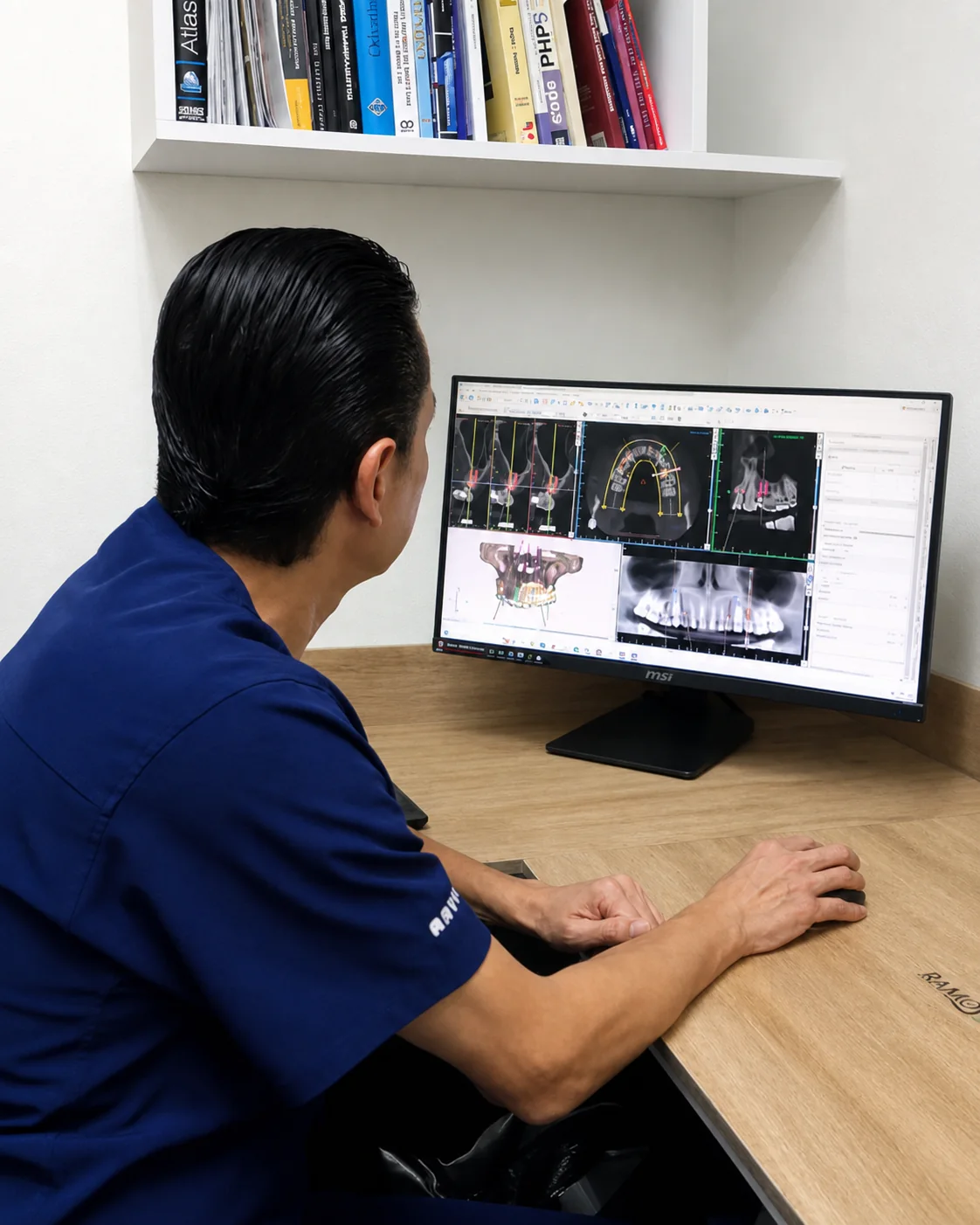
Planning software allows the available records to be reviewed together and aspects such as position, angulation and depth to be studied virtually before defining the clinical plan.
The image demonstrates the planning technology. The definitive recommendation depends on an in-person examination and the studies indicated for each patient.
04 · Guided surgery
When indicated, a customized guide can help transfer references from digital planning to the implant-placement procedure.
Guided surgery does not replace clinical judgment and is not necessary or appropriate in every case.
The following image shows a clinical procedure
It may contain a sensitive surgical view. You decide whether to reveal it.
05 · Design and manufacturing
With CAD technology, we design the restoration with form, fit and function in mind. The CAM process allows the piece to be manufactured by milling within a coordinated workflow.
Choose a stage to view digital design or milling. Videos play without sound and pause when they leave the screen.
The digital model allows the available anatomy to be studied and the restorative proposal to be developed before manufacturing.
These images and videos explain tools used in the digital workflow and do not represent a diagnosis, an individual treatment recommendation or a promised result. The appropriate technology and sequence must be confirmed during clinical evaluation.
Plan before you travel
Your existing photographs or X-rays can help the team understand your concern and prepare a preliminary review before your visit.